

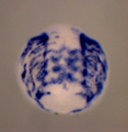
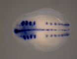
top left: expression of a muscle marker gene in developing Helobdella embryo (photo by D.-H. Kuo); top center: a Helobdella embryo labeled with different fluorescent lineage tracers in different cell lineages (photo by D.-H. Kuo); top right: expression of a cytoplasmic intermediate filament gene marking neuropile glia and nephridia in a Helobdella embryo (photo by D.-H. Kuo).
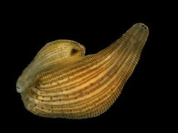
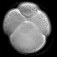
middle left: adult Helobdella robusta (photo by A. Rivera, edited by D.-H. Kuo); middle: an 8-cell Helobdella embryo (photo byD.-H. Kuo);
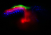
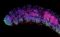
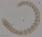
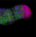
bottom left: confocal image of the head region of an enchytraeids worm (photo by S.-H. Chen); bottom center: bright-field image of an enchytraeid worm (photo by S.-H. Chen); bottom right: confocal image of blastema tissue (red) in a regenerating Enchytraeid worm (photo by S.-H. Chen);
Sizes are not in proportion.